Dr. Manasi Thakur | Gynaecologist In Nagpur | Pregnancy, Delivery | Abortion | Cosmetic Gynecology
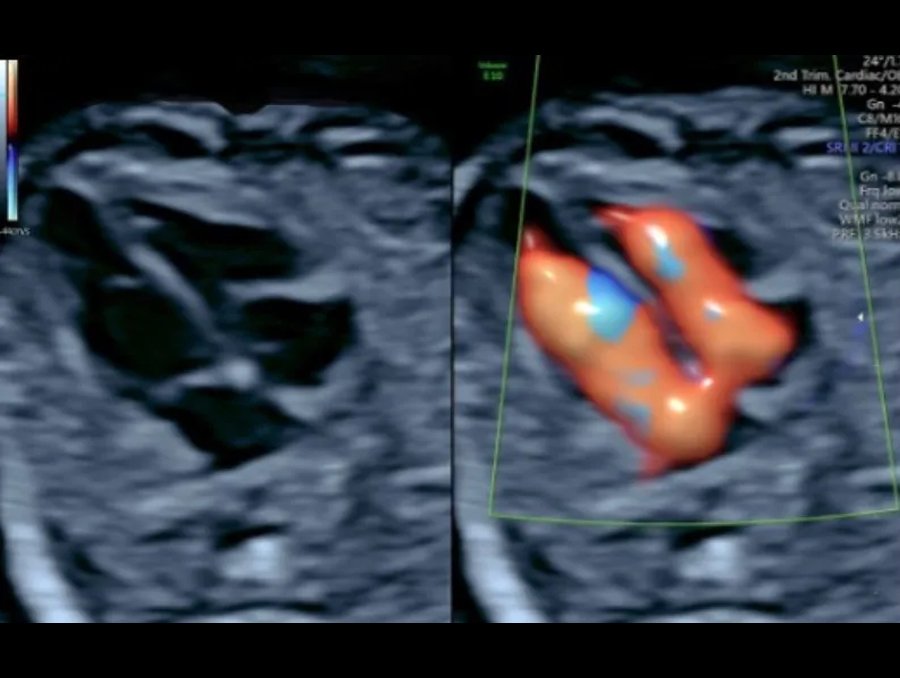
What is Fetal Echo?
When is Fetal Echo Recommended?
A fetal echo may be advised if the unborn child is at risk of heart abnormalities or related conditions, such as:
Family history of congenital heart disease
Previous child with a heart condition
Maternal use of alcohol or drugs during pregnancy
Use of certain medications (e.g., epilepsy or prescription acne drugs) that may affect the baby’s heart
Maternal medical conditions such as rubella, type 1 diabetes, lupus, or phenylketonuria (PKU)
How is the Test Performed?
Fetal echo at Zenith Hospital, Nagpur, under the care of Dr. Manasi Thakur, can be performed in two ways:
Abdominal Echocardiography – Similar to a regular ultrasound, where a probe is placed on the abdomen to capture images.
Transvaginal Echocardiography – The probe is gently inserted into the vagina, usually recommended in the early stages of pregnancy for clearer imaging.