Dr. Manasi Thakur | Gynaecologist In Nagpur | Pregnancy, Delivery | Abortion | Cosmetic Gynecology
Ultrasound in Pregnancy – Sonography Test Center in Nagpur | Pregnancy Sonography Price & Packages
All pregnancy sonography tests at Zenith Hospital, Nagpur are conducted strictly under PCPNDT laws. We strongly discourage and oppose the illegal practice of sex determination of the fetus.
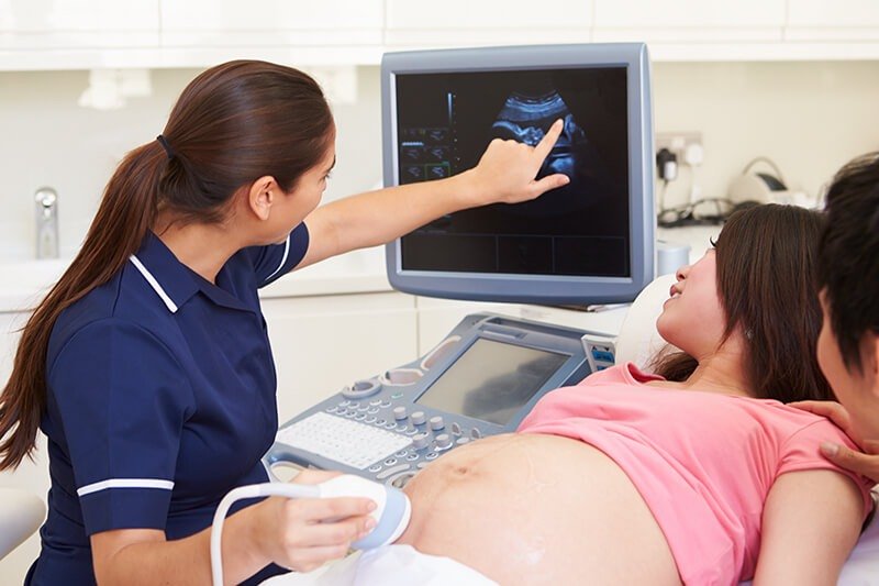
At Zenith Hospital, Nagpur, under the care of Dr. Manasi Thakur, we provide professional 3D and 4D pregnancy ultrasound services. With the latest technology in baby scanning, parents can view lifelike images of their baby and take home cherished keepsakes of this special journey.
Why Choose Zenith Hospital, Nagpur?
Sonography Tests Available at Zenith Hospital, Nagpur
We offer a wide range of ultrasound tests for pregnancy and women’s health, including:
Whole abdomen sonography
Pelvic sonography
Transvaginal sonography
Follicular monitoring
Fetal well-being scan
NT scan
Anomaly scan
3D/4D sonography
What is Sonography?
Sonography is a diagnostic imaging technique that uses high-frequency sound waves to create images of internal tissues and organs.
It is used to examine the abdomen, breasts, reproductive organs, blood vessels, and heart.
During pregnancy, it helps determine the age, number, position, and growth of the fetus and can also detect potential birth defects.
Beyond pregnancy, sonography can detect conditions such as gallstones, liver disease, or abnormalities in other internal organs.
Reasons for Sonography
Your doctor may recommend a sonography if you:
Experience pain, swelling, or symptoms of an internal organ disorder
Require monitoring of fetal growth during pregnancy
Need evaluation of the surgeon’s precision during procedures (such as biopsies)
It can also be used to examine:
Ovaries
Kidneys
Pancreas
Spleen
Testicles
Blood vessels
Gallbladder
Brain (in infants)
Uterus
Liver
Bladder
Eyes
Thyroid
Process of Sonography
A device called an ultrasound transducer is used to send sound waves into the body.
These sound waves bounce back as echoes, which are processed into images on the ultrasound machine.
The sonographer gently presses the transducer on the area being examined, and the images are used by doctors for assessment.
Steps include:
Performing an initial transvaginal ultrasound (when required).
Using safe imaging techniques for internal evaluation.
Repeating scans when needed for accurate monitoring.
Results of Sonography
Sonography allows doctors to:
Assess the health of organs, vessels, and tissues
Monitor the growth and well-being of the fetus
Identify abnormalities with high accuracy
Key Benefits
Safe, painless, and non-invasive procedure
No radiation exposure – uses sound waves
Safe for both mother and baby
Helps detect fetal growth issues early
To date, there is no evidence that ultrasound imaging causes harm to the fetus. It remains the safest method for monitoring pregnancy and other internal conditions.